A Digital (R)evolution in Palaeontology
Posted on 23 April 2013
A Digital (R)evolution in Palaeontology
 By Stephan Lautenschlager, Fellow and PhD Candidate at the University of Bristol
Palaeontology, the study of fossils and extinct organisms, has a reputation for being a very traditional and dusty discipline. Partly influenced by Palaeontologists’ portrayal in the media, the public imagines them roaming the badlands in the search for new fossils. Alternatively, they are seen as ivory tower scientists, spending most of their time in museum collections studying the bones of long extinct animals. Although both images hold a partial truth, palaeontology has experienced a tremendous paradigm shift in the last decade and has evolved into a multi-disciplinary science. Advances in hardware and software, and their wide and comparably inexpensive availability have led to a surge of computational methods to study fossils.
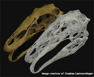
Non-destructive technologies, such as computed tomography (CT), used in combination with state-of-the-art visualisation techniques, allow the study, identification and, in the last step, visualisation of various fossil structures. For example, the brain and inner ear anatomy of dinosaurs, the bone microstructure of the first land animals and insects encased in amber.
Digital models can be used to simulate and evaluate locomotion or feeding behaviours. Computational methods, originally designed for the engineering industry, such as Finite Element Analysis, Multibody Dynamics Analysis or Computational Fluid Dynamics, are now finding their way into paleontological studies to investigate the functional and biomechanical behaviour of extinct organisms. In the course of the last decade, these methods have not only resulted in a plethora of new insights, but also created a veritable new sub-discipline, known as Virtual Palaeontology.
In spite of the wealth of information which can be gained from this brave new digital world, it also brings several difficulties with it.
Digitisation
Being computational techniques, these new methods all require a digital basis or template. This means that the fossils have to be digitised before they can be investigated. Although a variety of technologies are available to do so, they all come with advantages and disadvantages. For studies interested in internal structures of fossils or specimens still embedded in the rock, CT methods are the only possibility (apart from destructive sampling, which is rarely an option).
CT scanners are expensive, but have become more affordable for universities and large museums. If the necessary machinery is not available in the museum, many palaeontologists have found support in hospitals, which are often more than happy to use their medical scanners to scan a dinosaur skull instead of a human patient. Nevertheless, tomographic scanners are still not as widespread as one would hope, and scans are often expensive (up to £200-300 per scan).
An alternative is the 3D surface scanner, which uses visible or invisible laser light to capture the three-dimensional morphology of objects. These systems offer the advantage that they are capable of scanning a wide range of object sizes (from centimetre-sized ammonites to dinosaur skeletons), portable (meaning the scanner can be brought to the fossil, not the other way round) and relatively cheap. On the other hand, 3D scanners cannot capture the internal morphology (i.e. the internal form and structure of an organism). However, for many analyses the surface morphology is sufficient.
Recently a technique called photogrammetry has been employed. Photogrammetry produces three-dimensional models reconstructed from a series of two-dimensional photographs taken of an object. To reproduce the object geometry between 20 and 50 photographs, taken from different angles and positions, are computationally combined to produce the final model geometry. Photogrammetry maps texture information, included in the photographs onto the geometry, so that the photographed model is faithfully transferred into the digital realm. This has the obvious advantage that fossil specimens can be digitised in a timely, accurate and cost-effective manner. However, similar to other 3D surface scanning techniques, internal structures cannot be resolved.
Data formats
Apart from the availability of digitisation equipment, one of the biggest challenges the virtual palaeontologist faces, is the plethora of data formats, produced by digitisation methods, visualisation and analysis software. Digital models come in a variety of formats, including more common ones, such as .stl., .obj., .ply or .vrml, not to mention a range of proprietary formats which are only readable with specialised (and expensive) software packages.
Interchangeability between the individual formats is often limited and, if possible, can lead to other problems, such as the loss of information (e.g. colour, texture, coordinates) or a reduction of resolution.
Obviously, this complicates things for the individual researcher, trying to run different analyses, but also for institutions. The documentation of (fossil) specimens poses a considerable challenge to natural history collections and museums. Making specimens accessible to scientists and to the public is an essential but demanding task. The digitisation of selected specimens, or even whole collections, has been identified as a possible solution to this problem and many museums and universities are now employing 3D capturing techniques.
The resulting data can be made available via online repositories and different strategies are pursued to do so currently (such as Digital Morphology). However, the plethora of different data formats makes the dissemination difficult and, as of yet, there is no common standard or even agreement, which format would fulfil relevant criteria in terms of data access, sharing, completeness or sustainability.
Further reading
Bates KT, Falkingham PL, Hodgetts D, Farlow JO, B.H. B, O'Brien M, Matthews N, A., Sellers WI, Manning PL. 2009. Digital imaging and public engagement in palaeontology. Geology Today 25:134-139.
Falkingham PL. 2011. Acquisition of high-resolution three-dimensional models using free, open-source, photogrammetric software. Palaeontologia electronica 15:1-15.
Thessen AE, Patterson DJ. 2011. Data issues in the life sciences. ZooKeys 150:15-51.

